Inductive Roles of DCs, T Effector Cells, and Treg Cells in Determining Anti-Inflammatory or Proinflammatory Responses to Commensal and Pathogenic Bacteria and Soluble Proteins

Shown in Figure15 is a schematic representation of how the interactions of DCs with T effector and Treg cells may be influenced by the responses to commensal bacteria in the steady state or to pathogenic bacteria during infection or inflammation. In the steady state, a subpopulation of DCs in the intestine (shown in yellow) may be conditioned by the epithelial-cell-derived factors and promote the differentiation of FOXP3 Treg cells and IgA-secretory B cells upon migration to the mesenteric lymph nodes.

Figure 15.Panel A: Schematic representation of the interactions of DCs with T effector and Treg cell responses to commensal bacteria in the steady state. Panel B: The anti-inflammatory or proinflammatory responses in the steady state or during infection or inflammation. In the steady state, this conditioning may also occur following the sampling of commensal bacteria or alternatively to self antigens or to proteins found in food products, resulting in responses that “silence” the immune response through mechanisms of tolerance that include Treg cells or the effects of IgA-associated immune exclusion. A small number of another subset of DCs (shown in orange) may also be recruited to mesenteric lymph nodes in the steady state that may have escaped conditioning and drive T helper populations toward Th1 or Th17 profiles. Since these cells exist in small numbers, they will not give rise to disease in the steady state, but could act as sentinels when pathogenic bacteria are encountered or produced in excess, as in the case where aberrant responses to soluble proteins give rise to disease symptoms, as in the case of food allergy (Chapter 18 in Immunology IV). [Reproduced with permission from Bellanti, JA (Ed). Immunology IV: Clinical Applications in Health and Disease. I Care Press, Bethesda, MD, 2012

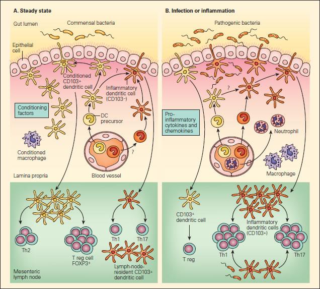
In contrast to the responses seen with commensal bacteria, some pathogenic bacteria possess sufficient virulence factors that allow them to invade the intestinal epithelium and to subvert the immune response and enhance their replication (Figure 15B). This would then result in the activation of the cytosolic- or cell membrane–associated activation of PRRs and the enhanced production of proinflammatory cytokines, e.g. TNF-α, and chemokines that would then promote the recruitment and influx of neutrophils, monocytes/macrophages, and DC precursors derived from the vascular compartment (Figure 15B). These newly arrived DCs have not undergone sampling of the luminal contents with conditioning; this sampling results in the production of Treg cells and IgA-associated immune exclusion, which then induce a cascading set of cellular responses brought about by proinflammatory Th1 and Th17 responses.


<<Back to Overview