HIV Virus
- HIV is a retrovirus and made of double stranded RNA enclosed within a glycosylated capsid
- Target cells for HIV are any cell expressing both CD4 and CCR5 or CXCR4 (chemokine receptors)
- HIV has evolved to use these receptors to infect the “central command” of the immune system and by so doing ultimately disables the immune response
- Infection is persistent and chronic and there is only one known case of someone being cured of HIV infection
- The prevalence of HIV (numbers of people infected) in sub-Sahara Africa vary from 6-30%, with southern Africa bearing the brunt of infections
- Understanding immunity to HIV is important for devising potential vaccine strategies as well as appreciating immunopathogenesis
HIV Infection
- Routes of HIV infection are predominantly through mucosal surfaces: male and female genital tracts, rectal surfaces and gut surfaces (perinatal infection)
- Acquisition of HIV can also be directly through the bloodstream from injection drug users
- Whatever the route of infection, there has been a defined stage of infection based on laboratory diagnosis
- Figure 1 shows Fiebig staging of laboratory testing for HIV infection.
- Fiebig staging is a 6-stage classification system that was formulated for staging early HIV infection based on the different times viral markers and host antibody responses emerge.
- The system was named after the paper’s first author
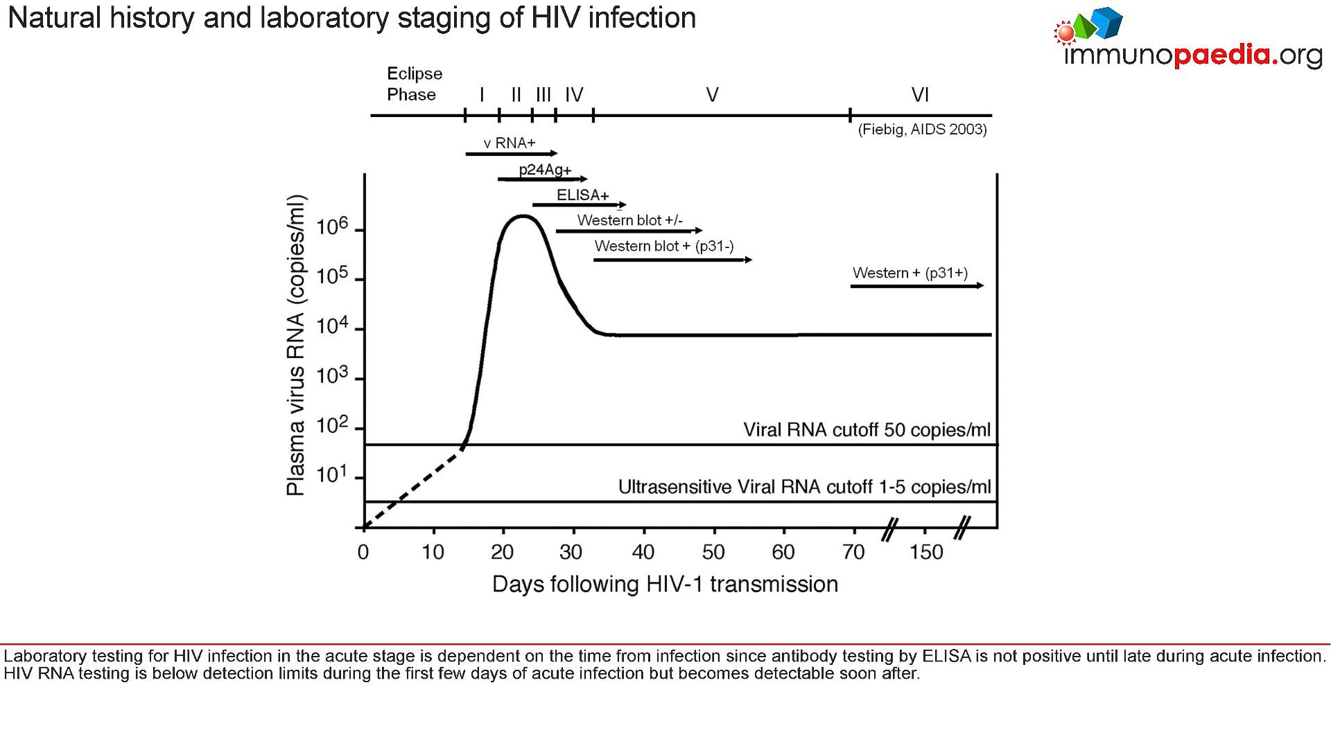
- For example, at the acute stage of HIV infection (Fiebig I/II), which is prior to seroconversion and at peak viraemia, there is a cytokine storm
- This is when a number of proinflammatory cytokine levels are high, giving rise to fever, pharyngitis,and lymphadenopathy
- As the peak viral load equilibrates to a set point, the ELISA test shows the presence of anti-p24 antibodies and production of large amounts of viral proteins. These are detected in western blots.
- The viral set point is usually established around 3-6 months after initial infection
- The set point is where the level of HIV replication in the host is relatively constant over time
- It is thought that this represents an equilibrium between host immunity to HIV and the rate of viral turnover
- What is important to realise is that HIV is predominantly found in lymphoid tissue and not in the blood circulation
- However, it is known that the higher the viral load in the blood plasma, the faster disease progression occurs
- Conversely, the lower the viral load, the slower disease progresses
- In some individuals, HIV infection appears to be under control, and it is in these individuals where biomarkers of immune control may be the most informative
The immune response to HIV
- There appears to be an ordered series of events that occur upon HIV acquisition
- Figure 2 shows a typical immune response to untreated HIV infection.
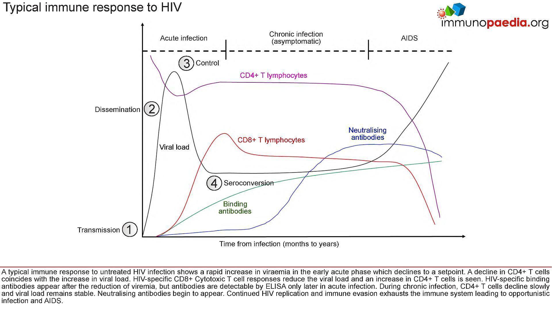
- After transmission of HIV to a new host:
- (1), there is dissemination of the virus to lymphoid tissues
- (2) a rapid increase in viraemia in the acute phase (measured as Fiebig stage I). The fall in peak viraemia is thought to be due to the initial immune control
- (3) and viral load declines to a set point.
- A decline in CD4+ T cells coincides with the increase in viral load.
- HIV-specific CD8+ Cytotoxic T cell responses are thought to reduce systemic viral load and an increase in CD4+ T cells is often observed.
- HIV-specific binding antibodies appear after the reduction of viraemia, but antibodies are detectable by ELISA only later in acute infection (4, Fiebig stage III onwards).
- During chronic infection, CD4+ T cells decline slowly and viral load remains relatively stable.
- Neutralising antibodies begin to appear only after about 3-6 months and continued HIV replication
- Immune evasion exhausts the immune system leading to opportunistic infection and AIDS.
HIV Transmission
- Infection is a “rare” event.
- In 80% of cases, transmission is thought to be established by a single virus
- All microorganisms that penetrate the epithelial surfaces are met immediately by cells and molecules that can mount an innate immune response (Figure 3)
- Epidermal Langerhans’ cells are a subset of dendritic cells found in the squamous epithelium of the female vagina and male inner foreskin and are the first immune cells to contact HIV during heterosexual contact.
- They express surface CD207 (langerin) that captures virus by binding to gp120, which induces internalisation and degradation of virus particles.
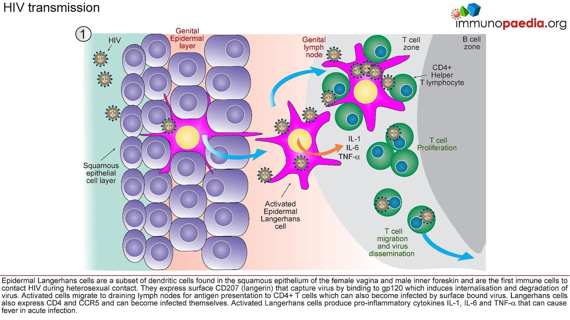
- Activated Langerhans’ cells migrate to draining lymph nodes for antigen presentation to CD4+ and CD8+ T cells.
- In the process, CD4+ T cells can also become infected by virus bound to the Langerhans cell surface (trans-infection).
- Langerhans’ cells may also express CD4 and CCR5 and can become infected themselves.
- Activated Langerhans’ cells produce pro-inflammatory cytokines IL-1, IL-6 and TNF-α that can cause fever.
- Dilation and increased permeability of the blood vessels during inflammation leads to increased local blood flow
HIV Dissemination
- Afferent lymphatic vessels drain fluid from the tissues and carry antigen bearing cells from infected tissues to the lymph nodes where they are trapped (Figure 4)
- Follicles expand as B lymphocytes proliferate to form germinal centres and the entire lymph node enlarges (lymphadenopathy)
- HIV infected CD4+ T cells, activated in genital draining lymph nodes, migrate to mucosal tissues such as the gut and skin.
- Dissemination of virus results in increased viral replication, mainly in lymphoid organs and leads to high viral loads in peripheral blood.
- There is also a rapid depletion of CD4+ T cells, particularly in the gut lymphoid tissues.
- Tissue macrophages express CD4 and CCR5 receptors and also become infected.
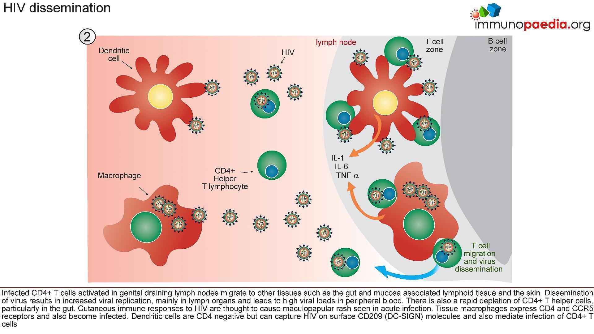
- Dendritic cells are CD4 negative but can capture HIV via surface CD209 (DC-SIGN) molecules and mediate trans-infection of CCR5-bearing CD4+ T cells
Control of Viraemia
- The partial resolution of peak viral load observed during the acute stage of HIV infection is associated with robust T cell immunity (Figure 5).
- Tissue dendritic cells engulf virus detected in extracellular spaces and present viral peptides by both HLA class I and II molecules in the lymph nodes to CD8+ and CD4+ T cells, respectively.
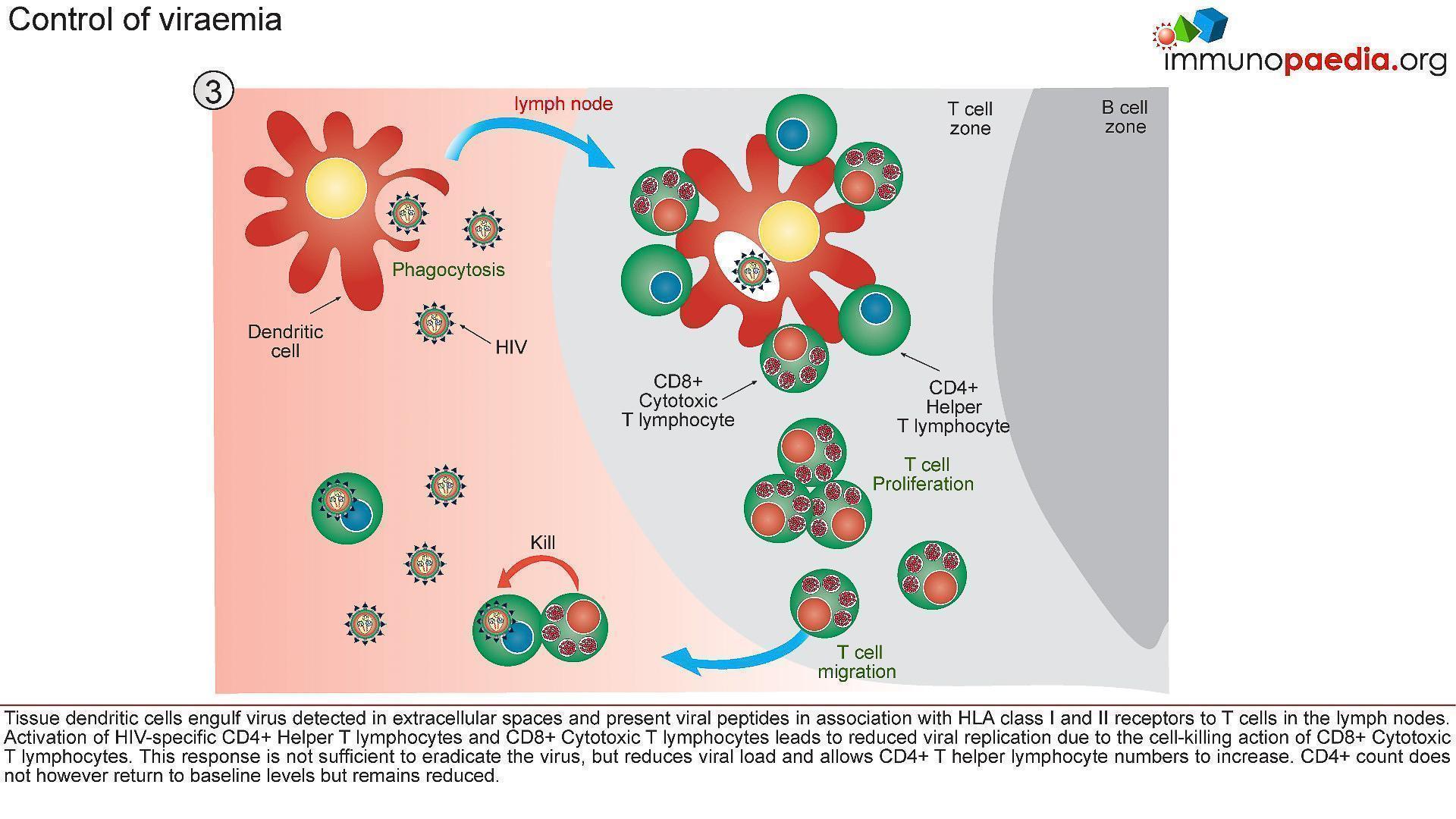
- Activated HIV-specific CD8+ cytotoxic T lymphocytes impart viral control by killing HIV infected cells and reducing viral replication.
- This response is not sufficient to eradicate the virus, but reduces viral load and allows CD4+ T helper lymphocyte numbers to increase.
- The absolute CD4+ count does not however return to baseline levels but remains reduced.
Seroconversion
- A multitude of immunological events have occured prior to seroconversion, many of them resulting in the clinical symptoms of acute retroviral syndrome.
- Antibodies to HIV (seroconversion) only begin to appear in peripheral blood 4-6 weeks after transmission, but in rare instances can take up to 3 months.
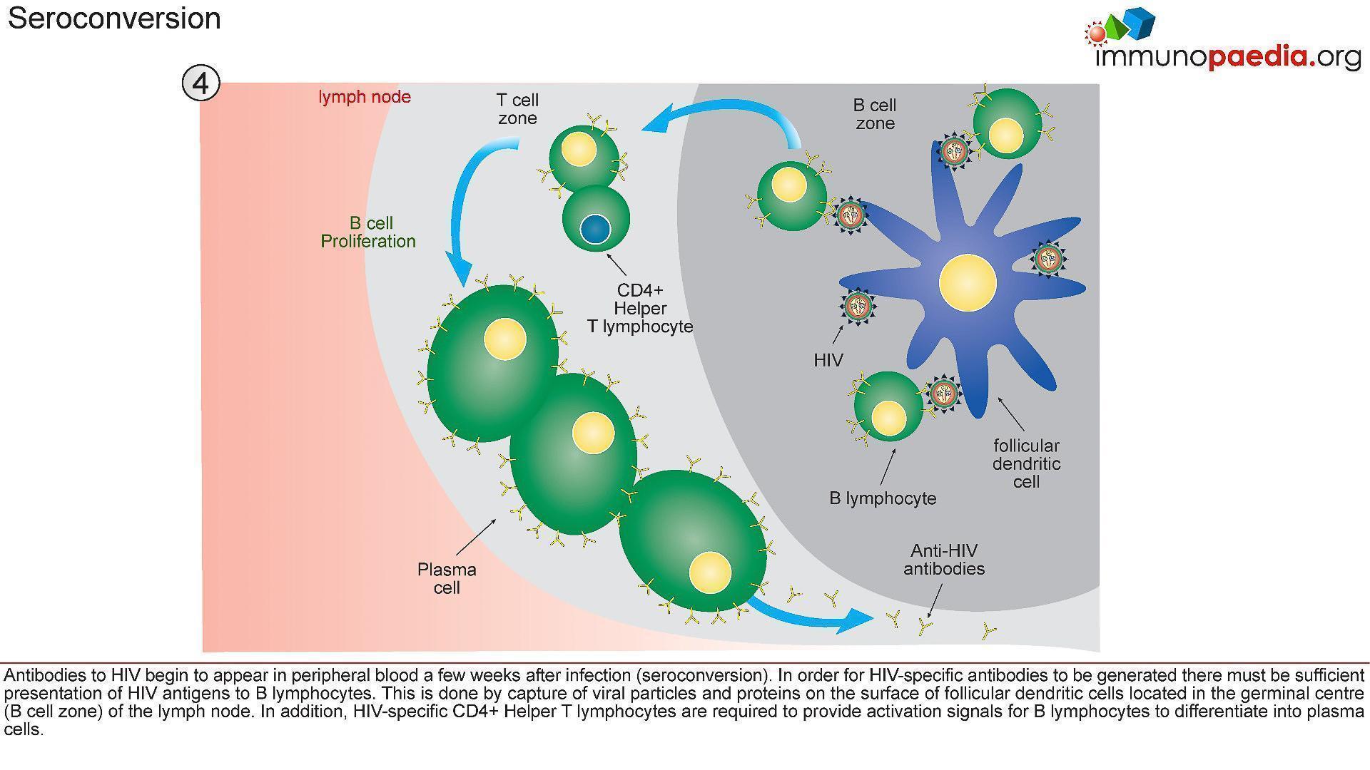
- In order for HIV-specific antibodies to be generated there must be sufficient presentation of HIV antigens to B lymphocytes (Figure 6)
- This is achieved by capture of viral particles and proteins on the surface of follicular dendritic cells located in the lymphoid follicles (B cell zone) of the lymph node.
- In addition, HIV-specific CD4+ helper T cells are required to provide activation signals for B cells to differentiate into plasma cells.
Quiz
Related Talk
Timothy Ray Brown, The Berlin Patient – The only known person to be cured of HIV
Wendy Burgers, University of Cape Town – Viruses and HIV
Associated Case Study
HIV Acute Retroviral Syndrome – A case of fever and general malaise
HIV Associated Dementia – Doctor, my sister is confused